Bachelor- und Masterarbeiten
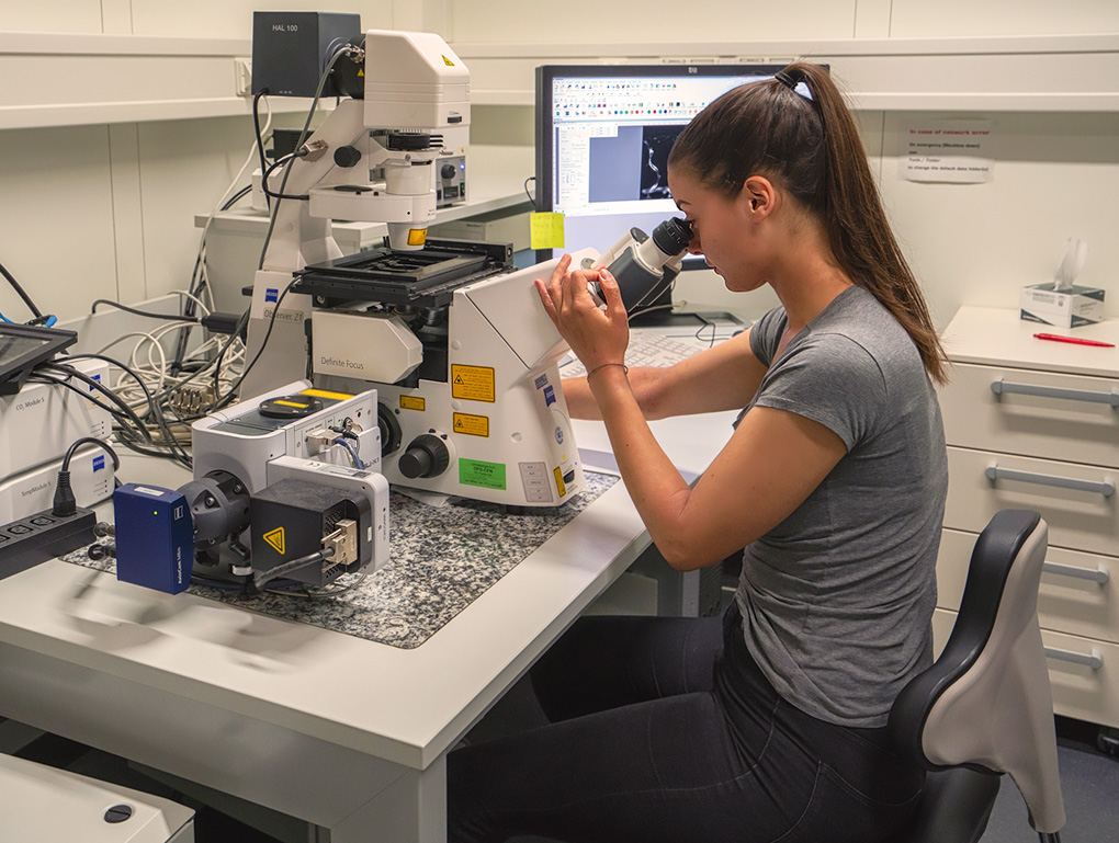
Mikroskopie Wir nehmen gern Studierende zur Durchführung von Masterarbeiten auf.
Die Inhalte, Methoden und Experimente der Arbeiten werden durch unsere Forschungsthemen und aktuellen Fragestellungen bestimmt. Interessenten/innen können sich jederzeit bei den Projektleitern/innen erkundigen.


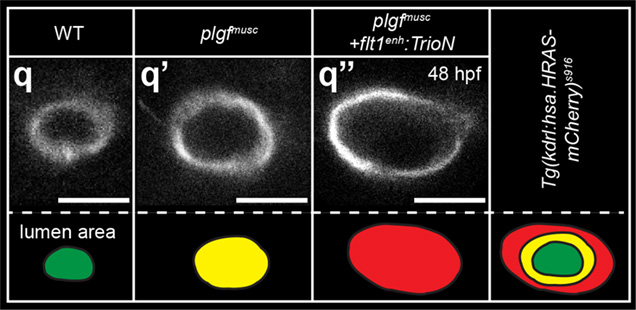
Visualization of arterial lumen diameter in Wildtype

Molecular targeting of arterial size: from zebrafish to clinical application

The arterial vascular network distributes blood flow through the body, a process crucial for sustaining organ function and homeostasis. During embryonic development arterial networks enlarge and expand to meet the increasing metabolic demand of the growing and differentiating tissue. Also in ischemic cardiovascular diseases, revascularization and regeneration (...)
Click here for detailed description of the Molecular targeting of arterial size Molecular targeting of arterial size: from zebrafish to clinical application

Background of your Master Project
The arterial vascular network distributes blood flow through the body, a process crucial for sustaining organ function and homeostasis. During embryonic development arterial networks enlarge and expand to meet the increasing metabolic demand of the growing and differentiating tissue. Also in ischemic cardiovascular diseases, revascularization and regeneration of hypoperfused organs involve adaptations of the arterial system. Selective targeting of the arterial growth process, and in particular creating arteries with a structurally large diameter to enhance flow conductance and relief hypoxia, is considered therapeutically relevant for the treating patients with ischemic cardiovascular diseases. The molecular and cellular mechanisms controlling arterial diameter are only partly understood but involve interactions between blood flow and endothelial cells, and molecular signaling cascades controlling distinct endothelial cell behaviors during the vascular remodeling process. The aim of this project is to identify molecular regulators of arterial diameter and to explore possibilities to translate these findings toward new clinical therapies to treat people suffering from ischemic cardiovascular diseases (as occurs in stroke, myocardial infarct, and peripheral arterial occlusive disease).

Your Project – Experimental Approach
In this project you will be investigating the interaction between the Vegf signaling pathway – a central regulator of blood vessel development - and small Rho GTPases in the context of arterial lumen regulation. In particular, we will address the role of these molecules and signaling pathways in regulating the size and shape of the vascular endothelium and smooth muscle cells, the major cellular components of blood vessels. To this end we generated a series of zebrafish mutants in which we silenced components of the Vegf and the RhoGTPase pathway using CrisprCas9 approaches. We will investigate the vascular morphology of these mutants using in vivo confocal imaging approaches in transgenic embryos, analyze the transcriptomic changes using state of the art single cell sequencing and next generation sequencing (NGS) approaches substantiated by loss and gain of function approaches of interesting candidate genes and pathways both in vivo and in vitro using vascular cells of human origin.

Techniques
In vivo confocal & two photon imaging, designing CrisprCas9 targeting constructs & how to genetically ablate gene function, transgenesis in the zebrafish embryo model, computational analysis of sequencing data and Big Data sets (some knowledge of “R” or Python is welcome), general histological techniques like whole mount in situ hybridization, RNA scope and antibody staining, general cell culture techniques, scientific writing.

Interactions with the scientific community
Our group has regular group meetings and journal clubs during which we discuss the emerging scientific concepts and experimental approaches. For the translational aspects of our work, we closely collaborate with clinicians (mainly from cardiology and internal medicine department) of Munich and Heidelberg University. We furthermore participate in the monthly seminars of several European vascular biology societies and the German Center for Cardiovascular Research.

Literature for this research topic
Klems, A, …. Le Noble, F. (2020). The GEF Trio controls endothelial cell size and arterial remodeling downstream of Vegf signaling in both zebrafish and cell models.
Nature Communications Oct 21;11(1):5319. doi: 10.1038/s41467-020-19008-0.


More info about this project
Please contact Prof. Ferdinand le Noble or Anna Lischke
 
Visualization of arterial lumen diameter in Wildtype
Figure 1 Visualization of arterial lumen diameter in Wildtype (WT, left panel), upon treatment with the Flt1 ligand Plgf (Plgfmusc, middle panel), and when combining Plgf with activation of the RhoGTP ase pathway by the guanidine exchange factor Trio (Flt1enh:TrioN, right panel). Arterial Lumen area color coded (green, yellow and red). Note the significant almost 2.5 fold increase in limen diameter upon combined Plgf+Trio therapy. Adapted from Klems & le Noble. Nature Comms, 2020.


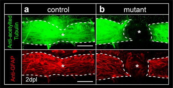
Visualization of spinal cord regeneration in control

In man, spinal cord injury induced destruction of neuronal tissues associates with disrupted motor- and sensory neuron signal processing, accounting for debilitating conditions. Repairing the damaged neuronal tissue, and restoring neuronal connectivity and function is considered of high medical relevance for treating affected patients. Unfortunately, in man neurons regenerate poorly and which molecular signaling pathways should be targeted (...)

Click here for detailed description of Novel pathways controlling Spinal Cord regeneration Novel pathways controlling Spinal Cord regeneration: from neural stem cell to functional motor neurons, zebrafish lead the way

Background of your Master Project
In man, spinal cord injury induced destruction of neuronal tissues associates with disrupted motor- and sensory neuron signal processing, accounting for debilitating conditions. Repairing the damaged neuronal tissue, and restoring neuronal connectivity and function is considered of high medical relevance for treating affected patients. Unfortunately, in man neurons regenerate poorly and which molecular signaling pathways should be targeted to restore regenerative capacity is an outstanding question in the field. In contrast to man, zebrafish show a remarkable regenerative capacity of both the central and peripheral nervous system. In the spinal cord, regeneration involves a complex interplay between neuron-derived cytokines like Vegf, blood vessels, oxygen radicals and the local inflammatory milieu controlling the behavior of macrophages and neutrophils. It is postulated that macrophages promote spinal cord regeneration by controlling post-injury inflammatory responses. However, the mechanistic basis of how macrophages regulate regeneration is poorly understood. To address this we generated a series of (tissue specific) mutants for neuron derived cytokines and their receptors, NADPH oxidases, as well as transgenics with a loss or gain of macrophage scenario and investigated repair processes upon spinal cord injury. We uncovered a hitherto unknown molecular mechanism involving precise control of neural growth factor dynamics, and interaction with the vascular system essential for controlling robust spinal cord regeneration in a spatio-temporal manner. We believe that these findings may translate into therapeutic strategies that may help to overcome neuro-degenerative diseases affecting motor neurons (like Amyotrophic lateral sclerosis, ALS) and improve motor neuron function upon spinal cord lesion.

Your Project – Experimental Approach
In this project you will be investigating spinal cord regeneration in a series of zebrafish mutants in which we silenced components of the Vegf, inflammatory and redox signaling pathway using CrisprCas9 approaches. We will investigate dynamic adaptive changes occurring at the neuro-vascular interface at the level of the spinal cord. We will do this using state of art imaging techniques that allow us to visualize cellular changes at single cell resolution. In addition we will use a series of transgenics that allow us to identify and cell-track individual neural stem cell, neural progenitor cells and their progeny including motor neurons. You will furthermore be involved in generating tissue specific mutants using CrisprCas9 approaches of molecules that we recently identified in transcriptomic screens. Finally, together with neuro-surgeons we will address to what extent the molecules identified in our screens, can be relevant in the human patient population.

Techniques
In vivo confocal & two photon imaging, designing CrisprCas9 targeting constructs & how to genetically ablate gene function in model systems, transgenesis in the zebrafish model, computational analysis of sequencing data, general histological techniques like whole mount in situ hybridization, RNA scope & antibody staining, scientific writing.

Interactions with the scientific community
Our group has regular group meetings and journal clubs during which we discuss the emerging scientific concepts and experimental approaches. For the translational aspects of our work, we closely collaborate with clinicians (mainly from the neurology and neurosurgery department) of the Charite (Berlin). We furthermore participate in the monthly seminars of several European vascular biology societies and the German Center for Cardiovascular Research.

Literature for this research topic
Cigliola, Becker, & Poss, K. (2020). Building bridges, not walls: spinal cord regeneration in zebrafish. Disease Models and Mechanisms. May 27; 13(5):dmm044131
https://www.ncbi.nlm.nih.gov/pmc/articles/PMC7272344/
DOI: 10.1242/dmm.044131


More info about this project
Please contact Dr. Andria Michael or Dr. Laetitia Preau
 
Visualization of spinal cord regeneration in control
Figure 1 Visualization of spinal cord regeneration in control (left panels) and gene mutant (right panels) scenario. Embryos were stained with antibody against acetylated tubulin to mark mature neurons (green), or with antibody directed against GFAP to mark glia cells (red). Note that control embryos regenerated the spinal cord lesion (lesion site indicated by *, regenerating area indicated by dotted line), whereas regeneration is absent in the mutant scenario.


Schematic representation of the RNA landscape

Information processing in the cell nucleus

Classically the genome has been divided in a coding part which contains most of the known genes and which is responsible for encoding the majority of the proteins known today, and into a so called non-coding part which was considered to be “junk DNA” and functionally not relevant. However, evidence is emerging showing that the non-coding area of the genome is in fact central to key biological processes and adaptive complexity in vertebrates. Non-coding RNAs (...)
Click here for detailed description of Information processing in the cell nucleus Information processing in the cell nucleus

Background of your Master Project
Classically the genome has been divided in a coding part which contains most of the known genes and which is responsible for encoding the majority of the proteins known today, and into a so called non-coding part which was considered to be “junk DNA” and functionally not relevant. However, evidence is emerging showing that the non-coding area of the genome is in fact central to key biological processes and adaptive complexity in vertebrates. Non-coding RNAs (ncRNA) such as micro RNAs, small nuclear RNAs, Piwi-interacting RNAs, and long-coding RNAs (lncRNA) have been shown to regulate gene expression networks by controlling nuclear architecture, transcription, mRNA stability and post-translational modifications, in a development and tissue specific manner. Especially the nervous system shows enriched expression of such regulatory ncRNA species. Dysregulation of ncRNA expression has been implicated in aging and age-related neurodegenerative disorders. More recently it was discovered that thousands of translated small open reading frames (sORF) exist in vertebrate RNA transcripts initially annotated as non-coding (ncRNA). sORFs generally lack sequence conservation and biological functions of encoded micro peptides remain hitherto largely unknown. Here we identified pan-vertebrate conserved sORFs. Using a combination of systems biology approaches including exploiting conservation signatures, Ribo sequencing, and proteomics approaches we identify a set of sORFs that could give rise to such micro peptides. At present the function of these micro-peptides and their molecular mechanism of action remain to be establish. We obtained evidence that some of these micro peptides may play a key role in regulating stem cell properties during development.

Your Project – Experimental Approach
In this project you will be investigating the role of non-coding RNAs and non-coding RNA- sORF derived micro peptides in neuro-vascular development. To this end we plan to generate a series of zebrafish mutants in which we silenced specific non-coding RNAs, and their micro peptide coding region using CrisprCas9 approaches. We will investigate the neuro-vascular morphology of these mutants using in vivo confocal imaging approaches in transgenic embryos, analyze the transcriptomic changes using state of the art single cell sequencing and next generation sequencing (NGS) approaches. We furthermore plan to establish human iPSC (induced pluripotent stem cell) derived organoids in which specific non-coding RNAs, or micro peptide thereof have been silenced.

Techniques
In vivo confocal imaging, designing CrisprCas9 targeting constructs & genetically ablate gene function, transgenesis in the zebrafish embryo model, computational analysis of transcriptome data and Big Data sets (some knowledge of “R” or “Python” is welcome), RNA scope, immune histochemistry, general cell culture techniques, working with induced pluripotent stem cell derived organoids, scientific writing.

Interactions with the scientific community
Our group has regular group meetings and journal clubs during which we discuss the emerging scientific concepts and experimental approaches. For the translational aspects of our work, we closely collaborate with clinicians (mainly neurologists) and systems biology experts specialized in addressing clinical issues.

Literature for this research topic
Chekulaeva and Rajewsky (2019). Roles of Long Noncoding RNAs and Circular RNAs in Translation. Cold Spring Harbor Perspect Biol. 2019 Jun; 11(6): a032680.
https://www.ncbi.nlm.nih.gov/pmc/articles/PMC6546045/


More info about this project
Please contact Prof. Ferdinand le Noble or Dr. Laetitia Preau
 
Schematic representation of the RNA landscape
Figure 1 Schematic representation of the RNA landscape. Left part (magenta), classical pathway, coding RNA, mRNA, protein. Right part (blue panels) overview of the different RNA species. Note non-coding RNAs containing small open reading frames (sORF) that can give rise to micro peptides. The project will investigate stem cell behavior in sORF and associated ncRNA loss and gain of function models.


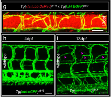
In vivo imaging of the spinal cord vascular network in the zebrafish embryo

Molecular regulation of angiogenic sprouting

The vascular network closely associates with the neuronal network throughout embryonic development, in adulthood and during tissue regeneration. Close association of vessels and nerves allows reciprocal cross-talk involving diffusible molecules, which is important for physiological functions in both domains. Arteries secrete factors that attract sympathetic axons, and adrenergic innervation of arteries allows (...)
Click here for detailed description of Molecular regulation of angiogenic sprouting Molecular regulation of angiogenic sprouting: organo-typical sprouting at the neuro-vascular interface

Background of your Master Project
The vascular network closely associates with the neuronal network throughout embryonic development, in adulthood and during tissue regeneration. Close association of vessels and nerves allows reciprocal cross-talk involving diffusible molecules, which is important for physiological functions in both domains. Arteries secrete factors that attract sympathetic axons, and adrenergic innervation of arteries allows the autonomic nervous system to control arterial tone and tissue perfusion. The nervous system on the other hand requires a specialized network of blood vessels for its development and survival. Metabolically active nerves rely on blood vessels to provide oxygen necessary for sustaining neuronal activity, and disturbances herein result in neuronal dysfunction. How nerves attract blood vessels is debated, but several studies addressing vascularization of the mouse embryonic nervous system suggest that the angiogenic cytokine VEGF-A is involved. In the mouse peripheral nervous system axons of sensory nerves innervating the embryonic skin trigger arteriogenesis involving VEGF-A - Neuropilin-1 (Nrp1) dependent signaling. While these studies provide evidence for the physical proximity and cooperative patterning of the developing nerves and vasculature, relatively little is known about mechanisms controlling VEGF-A dosage at the neurovascular interface. We recently identified a special form of angiogenic sprouting at the neurovascular interface. This project aims at identifying the molecular mediators of this special sprouting angiogenesis form. We are furthermore interested how such angiogenic sprouts subsequently remodel into a vascular network that innervates the spinal cord and how this process is regulated in a spatio-temporal manner. Finally we would like to investigate how changes in spinal cord vascularization affect the development of the peripheral nervous system, and if targeting the local vascular system can overcome neuro-degenerative processes as part of an effort to translate our experimental findings to the clinical setting.

Your Project – Experimental Approach
In this project you will be investigating how the interaction between blood vessels and the neural system shapes the spinal cord vascular network. We hypothesize that the Vegf signaling pathway may play a central role in the cross-talk between vessels and nerves. To address this hypothesis we generated a series of zebrafish mutants with a (tissue specific) Vegf gain of function scenario, or with a Vegf loss of function scenario using Tol2 transposome or CrisprCas9 techniques. We will investigate the vascular and neuronal morphology of these mutants & transgenics using in vivo confocal imaging approaches. We will furthermore perform transcriptomic and single cell sequencing approaches to identify potential cell-cell communication processes. This will be substantiated by loss and gain of function approaches of interesting candidate genes and pathways identified from the transcriptomic profiling using both the zebrafish embyo in vivo model system, and in vitro approaches in angiogenesis assays using vascular cells of human origin.

Techniques
In vivo confocal & two photon imaging, designing CrisprCas9 targeting constructs & how to genetically ablate gene function, transgenesis in the zebrafish embryo model, computational analysis of sequencing data and Big Data sets (some knowledge of “R” or Python is welcome), general histological techniques, RNA scope and antibody staining, scientific writing.

Interactions with the scientific community
Our group has regular group meetings and journal clubs during which we discuss the emerging scientific concepts and experimental approaches. For the translational aspects of our work, we closely collaborate with clinicians of Munich and Heidelberg University. We furthermore participate in the monthly seminars of several European vascular biology societies and the German Center for Cardiovascular Research.

Literature for this research topic
Wild, R, Klems, A, Preau, L.….le Noble, F. (2017). Neuronal sFlt1 and Vegfaa determine venous sprouting and spinal cord vascularization. Nature Communications, 8:13991. DOI: 10.1038/ncomms13991


More info about this project
Please contact Prof. Ferdinand le Noble or Dr. Laetitia Preau
 
In vivo imaging of the spinal cord vascular network in the zebrafish embryo
Figure 1 In vivo imaging of the spinal cord vascular network in the zebrafish embryo. Blood vessels are in green (using transgenic Tg(kdrl:eGFP) reporter line), neurons in red (using transgenic Tg(Xia.tubb:DsRed) reporter line). Upper panel (g): note the blood vessel network (green) surrounding the spinal cord neurons (red). Bottom left (h) trunk vascular network at day 4. Bottom right (i) trunk vascular network at 13 day of development. Note angiogenic sprouts (arrow heads) emanating from the trunk vessels at the level of the spinal cord. Adapted from Wild & le Noble, Nature Comms 2017.